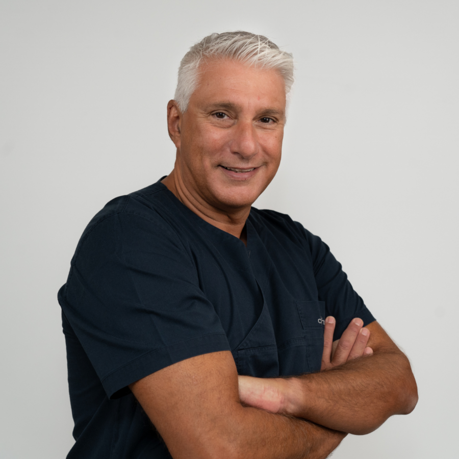
Presented by: Miltos Balidis MD, PhD, FEBOphth, ICOphth

Edited by: Penelope Burle de Politis MD

The surgical management of keratoconus has shifted over the last 20 years. Since the invention of intracorneal rings by Brazilian professor Paulo Ferrara and the development of corneal collagen cross-linking (CXL) by German professor Theo Seiler, advanced-stage keratoconus patients no longer rely on corneal transplantation as a sole hope for vision improvement. Given the pathophysiological basis of corneal ectasia – essentially the loss of collagen matrix biomechanical resistance, leading to stromal thinning and corneal deformity – it is understandable why tissue-adding approaches have taken the lead as keratoconus research progresses. While synthetic implants are still widely used with satisfactory results, real-tissue corneal implants such as CAIRS, conceived by Indian professor Soosan Jacob, have become the top trend.
As techniques continue to evolve, patients still await a cheaper, more accessible, quick-execution, fast-recovery procedure that does not depend on human donors. Maybe now they have it. Νamed after the ancient Greek concept of hospitality – ξενία – XENIA is a human-compatible collagen implant made from the purified extracellular matrix of porcine cornea. Following the example of other bioprosthesis, like the heart valves routinely used in cardiac surgery for decades, the XENIA material undergoes a biochemical decellularization process to completely devoid it from cells and molecular antigens. In comparison to other corneal procedures currently available for managing advanced keratoconus, XENIA has the advantages of being reversible, customized, rejection-free, tissue-additive rather than subtractive, and exchangeable – meaning it can be replaced anytime to align with eventual corneal changes.
The XENIA corneal implant for keratoconus consists of a 40 μm-thick, 8 mm-diameter circular disc of cross-linked – thus biomechanically strengthened – collagen. The lenticule is inserted into a specially designed pocket created with a femtosecond laser at a predetermined depth and with precise dimensions inside the cornea to increase its thickness. The implant enhances the biomechanical stability and architecture of the host corneal tissue while reshaping its aberrant anterior and posterior curvatures, ultimately improving visual acuity. The incision does not require stitches, as the cornea heals rapidly. Additionally, the technique can be combined with other therapeutic approaches, such as CXL, since the increase in corneal thickness facilitates CXL treatment.
The placement of the XENIA intrastromal implant is carried out under local anesthesia, without the need for hospitalization. The procedure is currently offered by only a few specialized centers worldwide, as it requires special equipment and excellent surgical training to do corneal delamination with maximum precision and at the right spot to achieve the best possible visual outcomes. For now, production of the intralaminar implants is limited and intended for patients with stage III and IV keratoconus who are on the waiting list for corneal transplantation. As with other types of intracorneal implants, the goal is to expand availability in the near future, even for earlier stages of the disease.
In this video, recorded in the main operating room of the Ophthalmica Eye Institute in Thessaloniki, Greece, Dr. Miltos Balidis (MD, PhD, FEBOphth, ICOphth), specialist in cornea and anterior segment ocular surgery, performs the first XENIA implantation in Greece on the left eye of a 45-year-old woman with advanced keratoconus, with the assistance of Egyptian professor and pioneer in XENIA implants Ahmed Elmassry. The surgical steps and timing in the video are as follows: corneal pocket creation with femtosecond laser (00:45); corneal pocket delamination (01:45); lenticule apprehension with forceps, ensuring correct side orientation (03:25); lenticule insertion (03:50); lenticule unfolding (04:05); intraoperative AS-OCT to verify proper lenticule placement without folds and away from the incision (06:35). The procedure ends with a successfully inserted XENIA corneal implant (07:09).
“Every problem has in it the seeds of its own solution.” – Norman Vincent Peale
Video:


